Cervical radiculopathy occurs when one or more nerves leaving the cervical spine become irritated or compressed. This commonly causes pain radiating from the neck into the shoulder, arm or hand and may be associated with numbness, tingling or weakness.
The two most common causes are an acute cervical disc prolapse (slipped disc) and age-related degenerative narrowing of the neural foramen (foraminal stenosis) causing nerve compression.
Although symptoms can be severe, many patients improve with non-operative treatment. Surgery may be recommended in carefully selected patients with persistent symptoms, progressive neurological deficits or significant functional impairment despite appropriate conservative management.
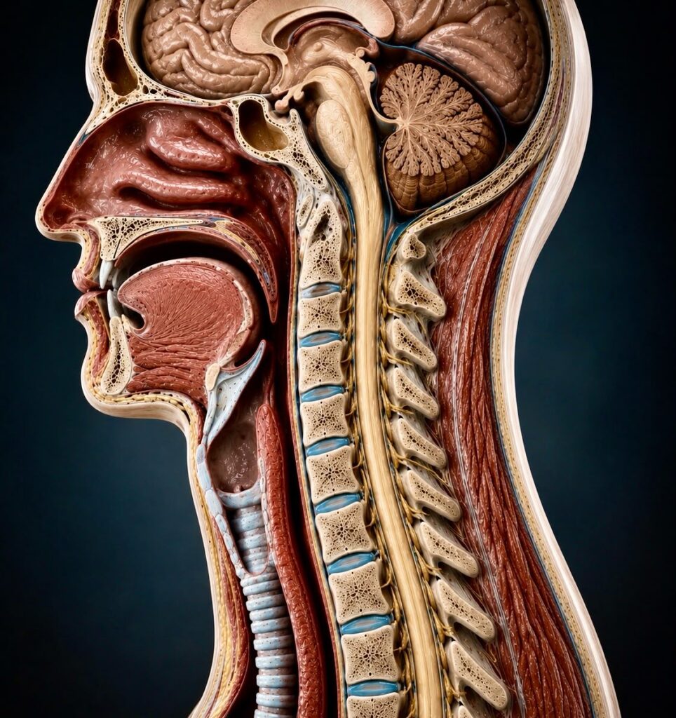
A normal cervical spine showing the spinal cord, intervertebral discs and exiting cervical nerve roots.
The cervical spine consists of seven vertebrae (C1–C7) separated by intervertebral discs. The spinal cord passes through the spinal canal, while individual spinal nerves leave the spine through small openings called the neural foramina.
Each cervical nerve supplies sensation and muscle function to specific parts of the shoulder, arm and hand. In a healthy spine, these nerves exit freely without compression.
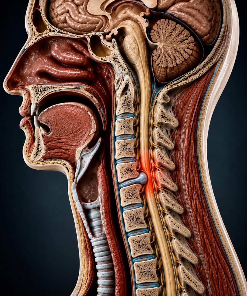
Acute cervical disc prolapse causing cervical nerve root compression.
Part of the soft inner material of the disc protrudes through the outer layer and compresses a nearby nerve root. This often affects younger or middle-aged adults and may occur suddenly following lifting or twisting, or occasionally without any obvious trigger.
With ageing, the intervertebral discs gradually lose height and elasticity. Bone spurs (osteophytes) and enlargement of the surrounding joints may progressively narrow the neural foramen through which the nerve exits. This is the most common cause in older adults.
Unlike cervical myelopathy, cervical radiculopathy affects a nerve root rather than the spinal cord. Symptoms are therefore typically confined to one arm and do not usually affect walking, balance or overall coordination.
Seek prompt medical assessment if you develop rapidly progressive weakness, significant loss of hand function or symptoms suggestive of spinal cord compression.
Diagnosis is based on a combination of the patient’s symptoms, neurological examination and imaging findings. MRI findings should always be interpreted alongside the clinical examination, as degenerative changes are common and do not necessarily cause symptoms.
Assessment of muscle strength, sensation, reflexes and nerve tension signs to identify the affected nerve root.
The preferred investigation for assessing the spinal cord, intervertebral discs and nerve roots.
Used when additional assessment of bone anatomy and osteophytes is required.
Flexion and extension views when spinal instability is suspected.
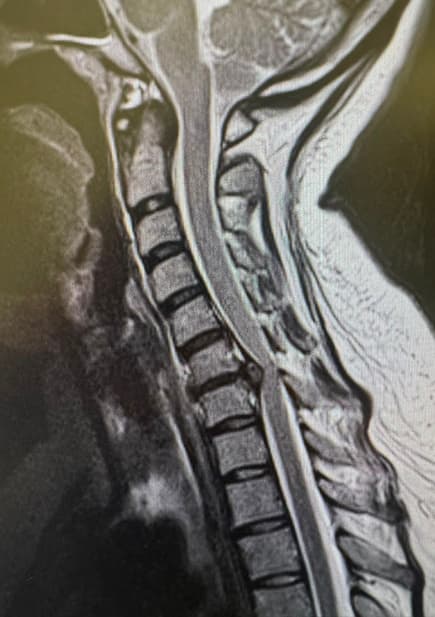
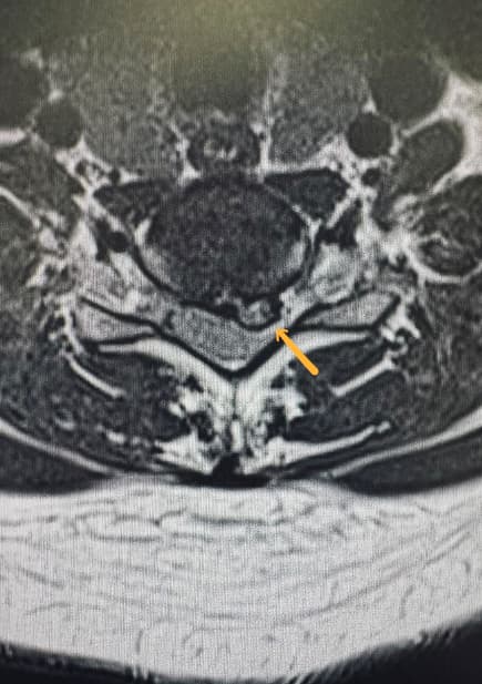
MRI demonstrating cervical nerve root compression caused by a disc prolapse.
The most appropriate procedure depends on the location of compression, the number of spinal levels involved, spinal alignment and patient-specific factors.
Arm pain remains severe despite an appropriate period of non-operative treatment.
Progressive muscle weakness and neurological symptoms develop.
Symptoms significantly affect daily activities or quality of life.
The choice of procedure depends on the underlying cause of nerve compression, the location of the pathology, spinal alignment and patient-specific factors.
Arm pain remains severe The most commonly performed procedure. an appropriate period of non-operative treatment.
A motion-preserving alternative in carefully selected patients.
Anterior decompression and stabilisation of the affected level.
Decompression from behind with stabilisation where indicated.
A motion-preserving procedure suitable for selected patients with isolated foraminal compression.
Occasionally indicated when compression extends behind the vertebral body or more extensive anterior decompression is required.
A motion-preserving alternative in carefully selected patients.
This information is intended for educational purposes only and reflects current evidence and accepted clinical practice at the time of publication. It should not be considered a substitute for an individual medical consultation. Treatment recommendations are personalised and should always follow assessment by an appropriately qualified spinal specialist.