Surgery is not required for every spinal condition. Many patients can be successfully managed through physiotherapy, rehabilitation, spinal injections, pain management and other conservative treatment strategies.
Where surgery is appropriate, the objective is to provide the safest and most effective treatment using the least invasive approach capable of achieving the desired outcome.
Mr Vittorio Russo offers the full spectrum of modern spinal surgical techniques, ranging from minimally invasive and motion-preserving procedures to complex spinal reconstruction, spinal tumour surgery, cranio-cervical junction surgery, deformity correction and advanced spinal oncology.
Every treatment plan is tailored to the individual patient, taking into account symptoms, neurological function, imaging findings, overall health, lifestyle and personal goals.
C1–C2 fusion is performed to stabilise abnormal movement or instability between the first and second cervical vertebrae.
Indications may include atlantoaxial instability, trauma, inflammatory disease, congenital abnormalities and selected degenerative or tumour-related conditions.
Modern techniques use specialised screws and rods to provide strong internal fixation while protecting the spinal cord, nerve roots and vertebral arteries. In complex cases, advanced imaging and intraoperative navigation may assist accurate implant placement.
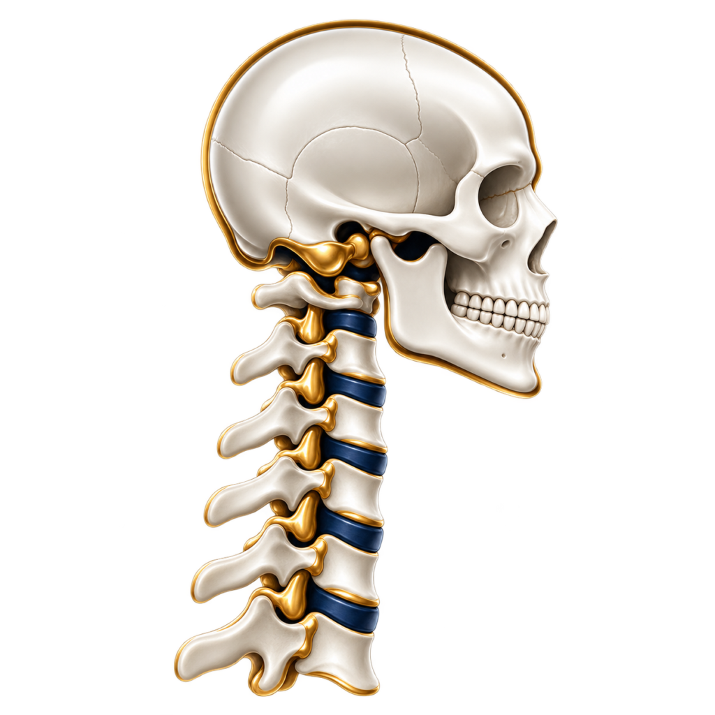
Occipito-cervical fusion stabilises the junction between the skull and upper cervical spine.
It may be required for severe cranio-cervical instability, basilar invagination, complex deformity, trauma, tumour, inflammatory disease or following previous surgery.
Specialised fixation connects the occipital bone to the upper cervical spine, with the extent of reconstruction determined by the underlying pathology.
Because of the important neurological and vascular structures within this region, these procedures require detailed pre-operative imaging, careful surgical planning and highly precise instrumentation.
Some cranio-cervical disorders require a combination of neural decompression, correction or reduction of deformity and stabilisation.
The surgical strategy is individually planned according to the relationship between the skull, upper cervical vertebrae, brainstem, spinal cord and vertebral arteries.
Complex reconstruction may incorporate C1–C2 fixation, occipito-cervical fusion or longer cervical constructs where required.
Anterior Cervical Discectomy and Fusion (ACDF) is one of the most commonly performed cervical spine procedures and is an established treatment for cervical radiculopathy and cervical myelopathy.
During the procedure, the diseased disc is removed to decompress the spinal cord and/or nerve roots. The disc space is then reconstructed using a specialised interbody cage or spacer positioned between the vertebrae to restore disc height, maintain alignment and promote fusion.
Depending on the patient’s anatomy and pathology, additional fixation may be achieved using integrated fixation systems or an anterior cervical plate secured with screws.
ACDF can provide effective neural decompression while restoring stability and maintaining appropriate cervical alignment.
Anterior cervical corpectomy is a more extensive anterior decompression used when compression of the spinal cord cannot be adequately addressed by removing the intervertebral disc alone.
The procedure involves removal of part or all of one or more vertebral bodies, together with adjacent discs where required, allowing direct decompression of the spinal cord. The resulting defect is reconstructed using a specialised cage or structural implant, usually combined with anterior fixation to restore stability and promote fusion.
Corpectomy may be considered for selected patients with severe cervical myelopathy, extensive spinal cord compression behind the vertebral body, ossification or calcification, tumour, deformity, trauma or complex multilevel pathology.
Cervical disc replacement is a motion-preserving alternative to fusion for appropriately selected patients with cervical disc disease.
The procedure uses an artificial disc prosthesis designed to preserve movement while restoring disc height and maintaining stability. Unlike fusion surgery, cervical disc replacement preserves movement at the treated level and may reduce mechanical stress on adjacent segments.
It may be suitable for selected patients with cervical radiculopathy and, in carefully selected cases, cervical myelopathy caused by cervical disc disease.
Posterior cervical decompression is frequently used for multilevel cervical spinal cord compression, cervical stenosis and complex cervical myelopathy.
The procedure involves removal of the posterior elements of the vertebrae to create additional space for the spinal cord. Where required, spinal stability is restored through instrumented fixation and fusion using screws and rods.
This approach can provide extensive spinal cord decompression while maintaining or restoring cervical alignment and long-term stability.
Cervical foraminotomy is a motion-preserving procedure designed to relieve pressure on an individual cervical nerve root.
It may be appropriate for selected patients with arm pain, numbness or weakness caused by nerve compression, particularly where decompression can be achieved without requiring fusion.
Laminoplasty is a motion-preserving spinal cord decompression procedure used in selected patients with multilevel cervical stenosis and myelopathy.
Rather than removing the posterior elements and performing a fusion, the procedure enlarges the spinal canal while aiming to preserve movement within the cervical spine.
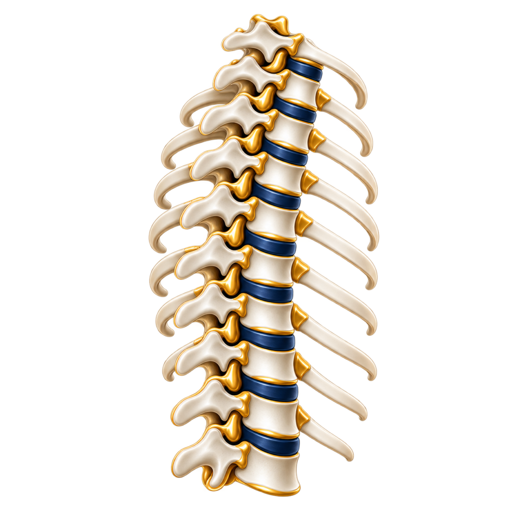
Posterior thoracic decompression is used to relieve pressure on the spinal cord caused by selected forms of spinal stenosis, ligamentous thickening, tumours and other compressive pathology.
Depending on the underlying condition, decompression may be performed alone or combined with instrumented spinal fixation and fusion when additional stability is required.
Thoracic disc herniation is relatively uncommon but can cause significant spinal cord compression and myelopathy.
The surgical approach is tailored to the location, size and characteristics of the disc herniation. Depending on the individual anatomy and pathology, approaches may include posterolateral or transpedicular techniques, costotransversectomy or an anterior approach through the chest.
Large, central or calcified thoracic disc herniations can represent particularly complex pathology and require detailed surgical planning to achieve adequate spinal cord decompression while minimising manipulation of the cord.
Thoracic fixation and fusion may be required for instability, fractures, deformity, tumours or following extensive decompression.
Modern techniques use specialised screws, rods and navigation technologies where appropriate to restore stability, protect neurological structures and reconstruct the spine.
Costotransversectomy provides access to the lateral and anterior regions of the thoracic spinal canal without requiring a conventional open thoracotomy.
It may be used for selected thoracic disc herniations, tumours and other complex pathology requiring access to the front or side of the spinal cord.
For selected complex conditions, particularly large central or calcified thoracic disc herniations, tumours or other anterior spinal pathology, direct access to the thoracic spine through the chest may provide the most appropriate route for spinal cord decompression.
Lumbar microdiscectomy is one of the most frequently performed spinal procedures and is used to treat sciatica caused by lumbar disc herniation.
Using microsurgical techniques, the portion of disc compressing the affected nerve is removed while preserving as much normal spinal anatomy as possible.
The principal objective is to relieve nerve compression and improve leg pain and associated neurological symptoms.
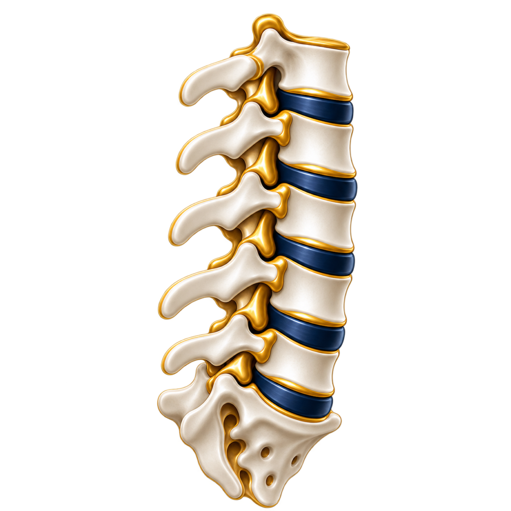
Lumbar decompression surgery is commonly performed for lumbar spinal stenosis causing leg pain, numbness, weakness or difficulty walking.
The procedure creates additional space for the affected nerves by removing the structures responsible for compression while preserving normal anatomy wherever possible.
Depending on the pathology, decompression may involve a laminectomy, laminotomy, lateral recess decompression and/or foraminotomy. In appropriately selected patients, these procedures may be performed using minimally invasive techniques.
Lumbar foraminotomy enlarges the opening through which an individual spinal nerve exits the spine.
It may be performed for foraminal stenosis causing radicular leg pain, numbness or weakness and can be undertaken alone or as part of a more extensive decompression or fusion procedure.
Lumbar fusion may be recommended where spinal instability, deformity, spondylolisthesis, recurrent nerve compression or other degenerative conditions require stabilisation of the spine.
Modern fusion surgery can be performed using several approaches, selected according to the individual patient’s anatomy, pathology and spinal alignment.
Posterolateral fusion is a well-established stabilisation technique using pedicle screws, rods and bone graft to achieve long-term fusion between vertebrae.
It is frequently used for instability, degenerative conditions, deformity and selected revision procedures.
TLIF combines nerve decompression with reconstruction of the disc space and stabilisation of the spine.
An interbody cage is inserted between the vertebrae and stabilisation is achieved using pedicle screws and rods.
TLIF may be used to treat spondylolisthesis, instability, recurrent disc disease, foraminal stenosis and selected degenerative or revision conditions.
ALIF approaches the lumbar spine from the front, through the abdomen, allowing placement of a relatively large interbody implant.
This can facilitate restoration of disc height, foraminal dimensions and spinal alignment while providing strong biomechanical support for fusion.
ALIF may be considered in selected patients with degenerative disc disease, spondylolisthesis, foraminal stenosis, spinal deformity and sagittal imbalance.
For complex spinal pathology, a combined anterior and posterior approach may be required to maximise stability, restore alignment and achieve robust reconstruction.
Lateral Lumbar Interbody Fusion, including the Extreme Lateral Interbody Fusion (XLIF) technique, approaches the lumbar spine from the side, allowing the damaged disc to be removed and replaced with a relatively large interbody cage.
This can restore disc height, improve spinal alignment and provide indirect decompression of the spinal nerves while minimising disruption of the muscles at the back of the spine.
Depending on the pathology, procedures may include microsurgical and tubular decompression techniques, mini-open approaches, percutaneous screw fixation, minimally invasive fusion procedures and hybrid reconstructive techniques.
Potential advantages in appropriately selected patients can include reduced tissue disruption, less postoperative discomfort, shorter hospital stay and earlier mobilisation.
The choice between minimally invasive and conventional surgery is determined by the underlying pathology rather than the size of the incision alone. The priority is always to select the approach that allows surgery to be performed safely and effectively.
Vertebral compression fractures may occur as a result of osteoporosis, trauma or tumour-related weakening of the spine.
While many fractures can be successfully managed with pain control, osteoporosis treatment, bracing and rehabilitation, selected patients continue to experience severe pain and functional limitation.
Vertebroplasty involves the percutaneous injection of specialised medical bone cement into a weakened or fractured vertebra.
Kyphoplasty uses a balloon or other specialised device to create a cavity within the affected vertebral body before bone cement is introduced.
In appropriately selected patients, the procedure can stabilise a painful vertebral fracture and may help restore some of the lost vertebral height.
Modern spinal surgery increasingly incorporates advanced technology to improve precision and support complex surgical planning.
Where appropriate, surgery may utilise computer-assisted navigation, robotic-assisted techniques, intraoperative imaging, advanced microscopy and intraoperative neurophysiological monitoring.
These technologies can be particularly valuable for complex instrumentation, revision surgery, deformity correction, tumour surgery and procedures involving anatomically challenging regions of the spine.
Technology remains an adjunct to surgical expertise, and its use is determined by whether it offers a meaningful advantage for the individual operation.
Extramedullary spinal tumours arise outside the spinal cord. They may be located within the spinal canal, or extend through the neural foramen into surrounding tissues.
For many intradural extramedullary tumours, surgery can be performed through a posterior microsurgical approach.
In cases of tumours extending also around the spine, treatment may require extended or occasionally also combined surgical approaches to safely address both the intraspinal and extraspinal components.
Where removal of the tumour or the surgical access required to reach it compromises spinal stability, instrumented spinal reconstruction and fusion may also be necessary.
Particularly complex tumours may require multidisciplinary planning involving spinal neurosurgery, spinal reconstruction and, where appropriate, thoracic, vascular or other specialist surgical teams.
Intramedullary tumours arise within the spinal cord itself and represent some of the most technically demanding conditions treated in spinal neurosurgery.
Surgery requires meticulous microsurgical technique because the tumour lies within functioning spinal cord tissue.
The spinal cord is carefully exposed and, where appropriate, opened through a precise microsurgical approach. The tumour is then separated from surrounding spinal cord tissue whenever a safe surgical plane can be identified.
Advanced intraoperative neurophysiological monitoring is used to assess spinal cord and neural function throughout surgery. The objective is to achieve the safest appropriate degree of tumour removal while placing preservation of neurological function at the centre of surgical decision-making.
Tumours arising from the vertebral column may cause pain, instability, deformity and compression of the spinal cord or nerve roots.
Treatment is highly individualised and may require tumour resection, spinal cord decompression and complex reconstruction using specialised implants.
Management is planned within a multidisciplinary framework and may be combined with radiotherapy, systemic treatment or other oncological therapies.
Cancer spreading to the spine can cause pain, vertebral collapse, instability and compression of the spinal cord.
Modern management requires close collaboration between spinal surgeons, oncologists, radiologists and radiotherapy specialists.
For selected patients, surgery may provide spinal cord decompression and stabilisation, preserve or restore neurological function, improve mobility and facilitate subsequent oncological treatment.
The extent of surgery is carefully tailored to the individual patient, their cancer diagnosis, neurological function, spinal stability and overall treatment objectives.
Thoracic and lumbar deformities include scoliosis, kyphosis and loss of normal spinal alignment.
They may result from degeneration, progression of pre-existing scoliosis, previous surgery, fractures, osteoporosis, trauma or other spinal conditions.
Patients may develop pain, postural imbalance, difficulty standing upright, reduced walking tolerance or neurological symptoms.
Surgery may involve decompression, interbody reconstruction, instrumented fusion and correction of spinal alignment, depending on the severity and complexity of the deformity.
Cervical deformity may cause progressive abnormal alignment of the neck, pain, neurological compression and difficulty maintaining a normal horizontal gaze.
Severe deformities, including chin-on-chest deformity, can interfere with vision, swallowing, mobility and daily activities. Reconstruction may require anterior, posterior or combined approaches, decompression, instrumented fusion and, in selected cases, osteotomies to restore appropriate alignment and stability.
Spinal deformity can disturb the normal balance of the trunk over the pelvis. Sagittal imbalance produces abnormal forward or backward posture, while coronal imbalance results in displacement of the trunk to one side.
When severe and symptomatic, treatment may require multilevel reconstruction to restore appropriate spinal alignment and balance.
Surgical planning considers the relationship between the spine and pelvis, neurological compression, symptoms and overall function rather than the degree of curvature alone.
Revision surgery may be required following previous spinal operations because of recurrent nerve compression, instability, non-union, implant-related problems, adjacent segment degeneration or progressive deformity.
Previous surgery can alter normal anatomy and create scar tissue, making revision procedures more complex. Treatment may range from targeted decompression to revision or extension of a fusion, replacement of implants or more extensive spinal reconstruction.