
Cervical myelopathy is caused by compression of the spinal cord in the neck. It commonly develops as a result of age-related wear and tear of the spine, although disc prolapse, inflammatory disease or tumours may also compress the cord.
Unlike a trapped nerve, which usually affects one arm, cervical myelopathy affects the spinal cord itself and may cause problems with hand function, balance, walking and coordination. Symptoms often develop gradually and may progress over time.
Early assessment by a spinal specialist is important, as prolonged compression of the spinal cord can result in neurological deficits that may become permanent if left untreated.
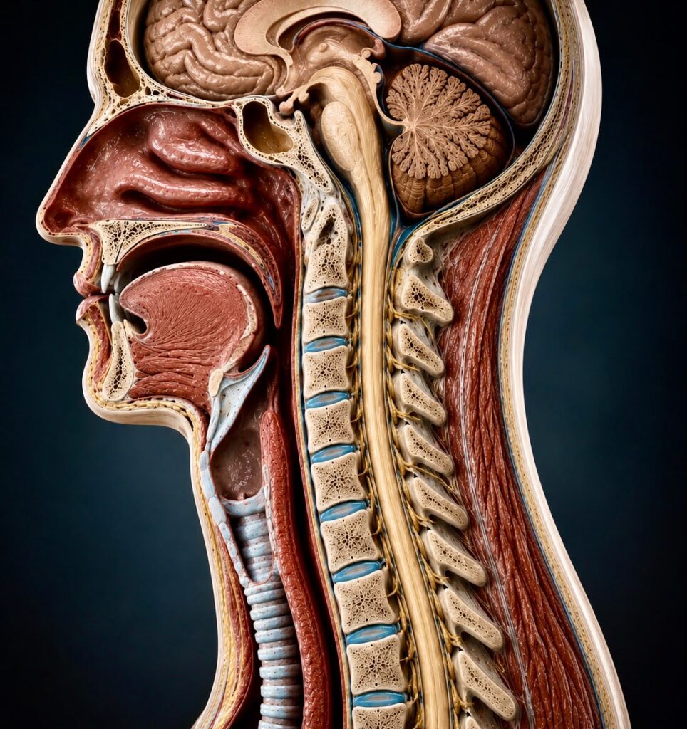
Normal cervical spine showing the spinal cord within a healthy spinal canal.
The cervical spine consists of seven vertebrae separated by intervertebral discs, which act as shock absorbers and allow movement of the neck. The spinal cord passes through a protective bony canal behind the vertebral bodies, with spinal nerves exiting at each level to supply the shoulders, arms and hands.
In a healthy spine, the spinal cord has adequate space within the spinal canal and is surrounded by cerebrospinal fluid, allowing it to function normally.
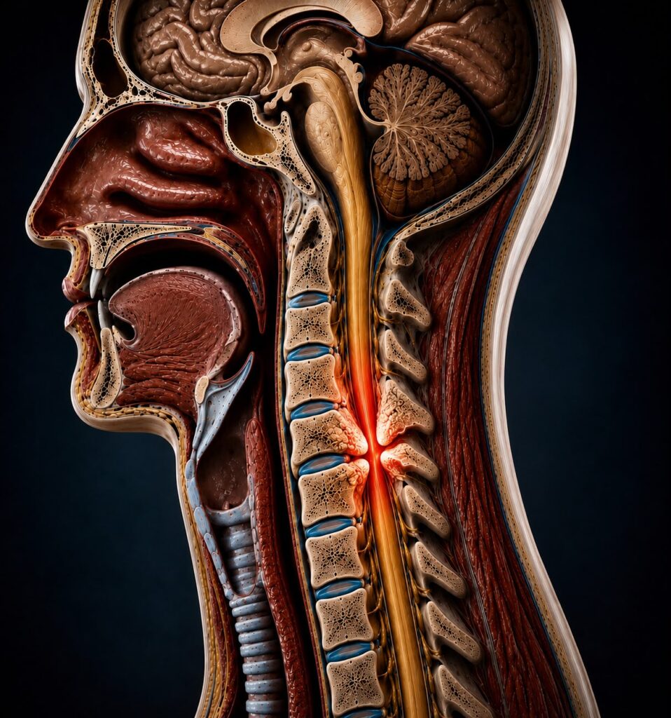
Cervical myelopathy showing compression of the spinal cord.
As we age, the structures of the spine gradually change. Intervertebral discs lose height, bone spurs (osteophytes) may develop, ligaments can thicken and the spinal canal may become progressively narrower.
When these changes reduce the available space for the spinal cord, pressure on the cord may develop. This can interfere with the transmission of signals between the brain and the rest of the body, leading to problems with movement, sensation and coordination.
The severity of symptoms does not always correspond directly to MRI appearances, which is why both clinical assessment and imaging are important when deciding on the most appropriate treatment.
Diagnosis is based on a combination of your symptoms, neurological examination and imaging findings. MRI findings are always interpreted alongside the clinical assessment, as imaging alone does not determine whether treatment is required.
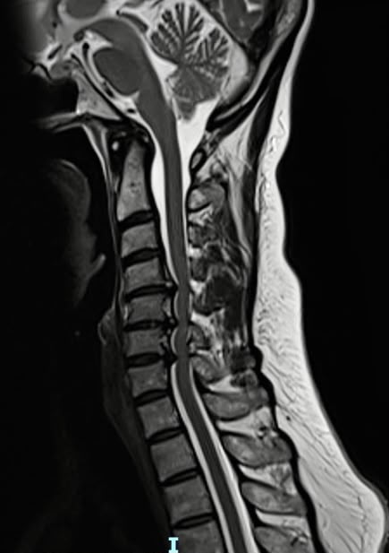
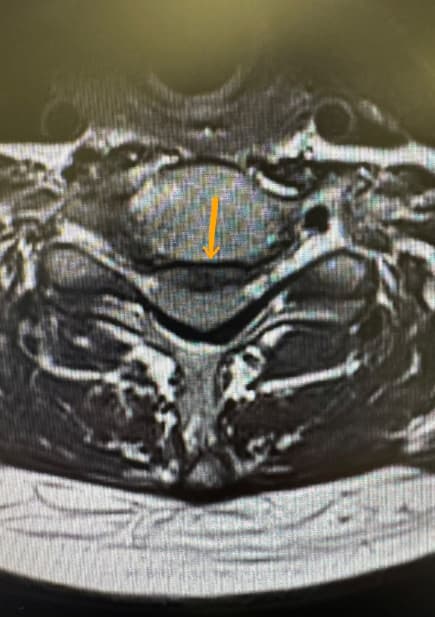
MRI scan demonstrating cervical spinal cord compression.
Assessment of strength, sensation, reflexes, coordination and balance, the findings that most often determine urgency.
The single most important investigation for evaluating spinal cord compression and the condition of the cord itself.
Used when additional detail about the bony anatomy, ossification or previous fusion is required.
Flexion and extension views when spinal instability is suspected or alignment must be assessed.
Treatment depends on the severity of symptoms, the neurological examination, MRI findings and overall health.
Patients with minimal symptoms and stable neurological function may be monitored with regular clinical review and repeat imaging when appropriate. Cervical myelopathy has a variable natural history: some patients remain stable for prolonged periods, while others deteriorate gradually. Prompt review is advised if hand function, balance, walking, limb strength or bladder control worsen.
Physiotherapy and other conservative measures do not relieve spinal cord compression, but may help maintain mobility, optimise general spinal health and manage associated symptoms in carefully selected patients. Conservative care should be supervised, with regular reassessment to confirm neurological function remains stable.
Where neurological symptoms are progressive or significant spinal cord compression is present, surgery may be recommended to relieve pressure on the cord, reduce the risk of further deterioration and optimise the opportunity for neurological recovery.
The most appropriate procedure depends on the location of compression, the number of spinal levels involved, spinal alignment and patient-specific factors.
Commonly used for one or two levels of anterior spinal cord compression.
A motion-preserving alternative for carefully selected patients.
Used when compression extends behind the vertebral body and more extensive anterior decompression is required.
Expands the spinal canal while preserving motion in selected patients with multilevel disease.
Suitable for many patients with multilevel myelopathy, particularly where instability or deformity is present.
Occasionally required for complex deformity, severe multilevel compression or revision surgery.
The information provided on this page is intended for general educational purposes only. It does not constitute medical advice and should not be used as a substitute for an individual consultation with an appropriately qualified healthcare professional. Treatment recommendations vary according to each patient’s medical history, symptoms, clinical examination, imaging findings and personal circumstances. If you have concerns about your health or develop new or worsening neurological symptoms, you should seek prompt medical assessment.